
3D X-Rays & Diagnostics in Pleasant Hill, CA
- Modern Technology
- Cosmetic Dentistry
- Personalized Care
- Crafted Smiles
- Natural Results
- Same-Day Solutions
Accurate Imaging for Better Treatment Decisions
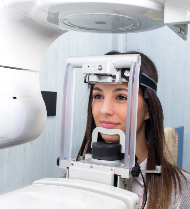
3D dental X-rays produce detailed three-dimensional images of the teeth, roots, jaw, and bone structures that conventional flat X-rays cannot capture, enabling more precise diagnosis and treatment planning for implants, extractions, orthodontics, and complex restorations. Advanced diagnostics reduce the guesswork in dental care and give both provider and patient a clear view of what is happening before any treatment begins. In a region where patients frequently arrive having seen multiple providers across Walnut Creek, Concord, and the wider Contra Costa corridor, starting with complete, high-resolution imaging means treatment recommendations here are built on evidence, not on another practice’s outdated records.
At Cosmetic Dental Spa Ricardo M. Perez, DDS, we offer dental 3D X-rays and advanced diagnostics in Pleasant Hill, CA, as a standard part of treatment planning for any procedure that benefits from them.
What Are 3D Dental X-Rays and Advanced Diagnostics?
Three-dimensional dental imaging, most commonly delivered through cone beam computed tomography (CBCT), captures a volumetric scan of the jaw, teeth, sinuses, and surrounding bone in a single rotation. Unlike a standard two-dimensional X-ray, which flattens overlapping structures into a single image, CBCT allows the provider to view anatomy in cross-sectional images from any angle. This is particularly important for procedures where millimeters matter, such as implant placement near a nerve or extraction of an impacted tooth.
The diagnostic suite at this practice also includes low-radiation digital X-rays, laser cavity detection, and an intraoral camera. Each tool serves a different purpose, and together they form a diagnostic baseline that supports accurate, evidence-based treatment decisions rather than clinical assumptions.
What Can 3D X-Rays and Dental Diagnostics Address?
Implant Planning and Bone Assessment
CBCT imaging maps bone density, volume, and proximity to nerves and sinuses with the precision required for safe, accurate implant placement. This level of detail is not available on standard X-rays.
Complex and Impacted Extractions
Root anatomy, position relative to the nerve canal, and surrounding bone structure are clearly visible in 3D, reducing surgical risk and improving outcomes for difficult extractions, including impacted wisdom teeth.
TMJ and Bite Evaluation
3D imaging provides a structural view of the temporomandibular joint, helping identify joint degeneration, disc displacement, or bony changes that contribute to jaw pain, clicking, or limited opening.
Early Cavity Detection
We use laser fluorescence to detect demineralization in enamel at a stage earlier than traditional dental X-rays or visual exams can detect, enabling conservative intervention before drilling is needed.
Who Are 3D X-Rays and Diagnostics For?
Patients being evaluated for dental implants who need bone volume and nerve proximity confirmed before surgery
Anyone with impacted or complex wisdom teeth requiring extraction planning
Patients experiencing jaw pain, clicking, or bite dysfunction that warrants TMJ structural evaluation
Adults undergoing orthodontic or clear aligner treatment where root positions affect the plan
Patients with a history of cavities who want early detection to catch decay before it progresses
Anyone preparing for a complex restorative case, such as full-mouth reconstruction or bridge planning
New patients establishing a comprehensive diagnostic baseline
When Are 3D X-Rays Not the Right Fit?
Advanced imaging is recommended based on clinical need, not as a default. Not every appointment or procedure requires 3D imaging, and the practice follows ALARA principles (as low as reasonably achievable) when determining whether the diagnostic benefit justifies the scan.
Routine cleanings and exams
Standard digital X-rays provide the information needed for preventive visits. CBCT is not required.
Small cavities or simple fillings
Two-dimensional imaging is adequate for diagnosing and planning standard restorations.
Cosmetic-only consultations
Veneer or whitening consultations do not typically require 3D imaging unless underlying structural concerns are present.
Pregnancy
Elective imaging is deferred during pregnancy as a standard precaution, though emergency imaging with proper shielding remains available if clinically necessary.
Our Diagnostic Imaging Process
Health History and Clinical Indication Review
Before any imaging is ordered, the clinical need is established through a conversation about your symptoms, goals, and dental history. The imaging type is selected based on what information is needed for accurate treatment planning.
Digital X-Rays and Diagnostics
Bitewing, periapical, or panoramic X-rays are taken as indicated. If needed, cavity screening is also performed.
Treatment Planning
Diagnostic results are integrated into a treatment plan tailored to your situation. If a specialist referral is appropriate, it is made with the imaging already completed, so the specialist has the information they need from the start.
Before and After Your Appointment
Before Your Appointment
- Remove any removable dental appliances, retainers, or jewelry around the head and neck before imaging.
- Let the team know if you are or may be pregnant so imaging decisions can be made accordingly.
- Bring any recent X-rays from a previous dentist if you are a new patient; this may reduce the need for repeat imaging.
After Your Appointment
- Diagnostic imaging involves no recovery; you can resume all normal activities immediately.
- Your provider will review findings with you during or after the appointment, using the images to illustrate what was detected.
- A copy of your X-rays can be provided on request for your records or for a second opinion.
Why Choose Dr. Perez for Dental Diagnostics?
Diagnostic tools are only as useful as the provider interpreting them. Dr. Perez’s training in cosmetic dentistry, jaw relations, and implant planning means findings are evaluated in the context of how teeth, bone, and bite function together.
Our practice philosophy emphasizes transparency. When imaging reveals a finding, patients see it on the screen and receive a plain-language explanation of what it means, the options, and what happens if nothing is done. For patients who have felt uncertain or uninformed at dental offices in the past, that clarity is essential.
Frequently Asked Questions
What is the difference between a regular dental X-ray and a 3D scan?
Standard X-rays show flat, two-dimensional images. 3D scans show bone, roots, and nerves in full spatial detail. Conventional X-rays are effective for detecting cavities and for routine monitoring.
Are dental X-rays safe?
Yes, digital X-rays use up to 90% less radiation than traditional film. The exposure from a full set of digital X-rays is comparable to what you receive during a few hours of normal background radiation. 3D CBCT exposes patients to more radiation than standard X-rays and is recommended only when the clinical benefit outweighs the risk.
How often do I need dental X-rays?
Frequency depends on your oral health history and the clinical questions being evaluated. Your provider will recommend a schedule based on your individual situation.
How much do 3D X-rays and diagnostics cost?
Cost depends on the type of imaging taken and the clinical need driving the recommendation. Factors affecting your total include the number of images, the complexity of the diagnostic question, and your insurance coverage for each imaging type.
Does Dr. Perez accept insurance?
Yes, standard digital X-rays are covered by most dental insurance plans as part of routine diagnostic care. Coverage for 3D cone beam CT imaging varies more widely and typically requires documented clinical necessity, such as implant planning or surgical evaluation, for reimbursement. The team will verify what your plan covers before any imaging is taken.
Good Treatment Starts With A Clear Picture
Cosmetic Dental Spa Ricardo M. Perez, DDS
Dentistry Powered by Innovation
3D DIGITAL IMAGING
Cone beam imaging captures detailed three-dimensional views of teeth, roots, and bone that standard X-rays cannot provide.
CEREC SAME-DAY CROWNS
Chairside CAD/CAM technology designs and mills permanent ceramic crowns in the office, completing the entire restoration process in a single visit without temporaries or lab waiting periods.
Intra-oral Scanning
Digital impressions replace the discomfort of traditional impression trays with a quick, precise 3D scan of the teeth, supporting planning and accurate treatment previews.
Diode Laser Technology
Laser-assisted procedures enable precise gum contouring, soft tissue treatment, and selected periodontal procedures with reduced bleeding and faster healing.